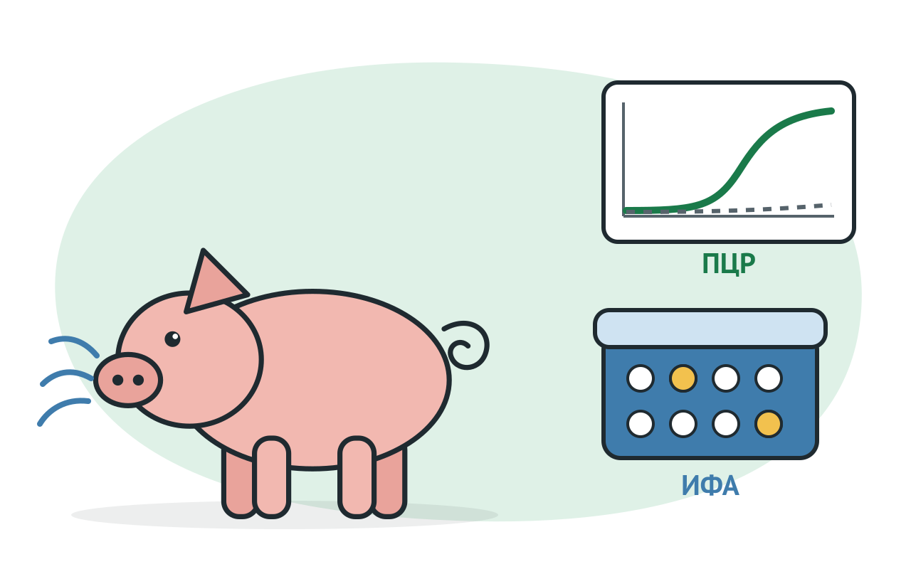

Pig respiratory pathology is described as a respiratory complex (porcine respiratorial disuse complex, PRDC). It's polymicrobial: the disease arises from different combinations of primary and secondary agents. The Opriesnig and co-authors review included the Pig Respiratory Syndrome Virus (RRS), pig influenza virus, Type 2 swine circulatory virus (PCB-2), Mycoplasma hyopneumoniae and Pasteurella multitocida.
Combinations change the severity of the disease. According to the same survey, co-infection between CBS-2 and M. Hyopneumoniae, which is separately low-pathogenic, causes severe respiratory disease.
The clinical picture doesn't breed these infections. The management of the World Organisation for Animal Health (WOAH), WOAH, clearly says that pig flu is indistinguishable by signs from RFDS, Auresca's disease, pork respiratory coronavirus, actinobacillosis pheurropneumonia and mycoplasmosis, so laboratory research is needed. So the practical conclusion is, a panel, not one test.
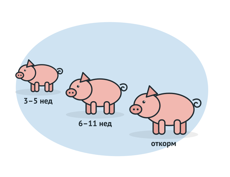
The group's age narrows down the list. In a 2021 study, tracheobronchial swine swine was swabbed with signs of respiratory disease in the households of Belgium and the Netherlands. Proportion of positive in M. Hyopneumoniae grew up with age: 8.5 per cent in 3–5 weeks, 16.2 per cent in 6–11 weeks and 53.4 per cent on board.
| Age | Which was found more frequently (% of positive samples) |
|---|---|
| 3-5 weeks | Influenza A virus: 28.4%, pig cytomegalovyrus: 18.2%, A. pleuropneumoniae: 24,7%, PRRSV-1: 11,5% |
| 6 - 11 weeks | A. Pleuropneumoniae: 32.1%, pig cytomegalovyrus: 26.7%, PRRSV-1: 25.5%, influenza A: 20.5%, CBS-2: 11.4% |
| Feeding | M. hyopneumoniae: 53,4% |
The most common pairs in one sample were RPTC and Micoplasmosis, RPTC and flu, RPTC and FMC-2, Micoplasmosis and influenza. Of the triple combinations, the GFMV + M prevailed. Hyopneumoniae + influenza A virus, as well as RPTC + M. Hyopneumoniae + CBS-2. This is a specific study, not a standard, but it's logical: before they leave, they test viruses first, and when they feed, they start with mycoplasmosis.
The task defines both material and method.
| Flash | Monitoring the herd | |
|---|---|---|
| Who are being explored | clinically ill and fallen animals | planned sample by age group |
| Sample type | Lungs, almonds, lymph nodes, blood, lower respiratory swabs | serum, oral fluid, tracheal swabs, meat juice |
| Method | PCR for the driver | ELISA on antibodies plus PCR randomly |
| What do you get? | confirmation of case | Seroprophylaxis and age of infection |
Serology in an acute situation helps the weak: antibodies appear later on. The PCR is also limited in routine surveillance: it only catches the driver at the moment of release.
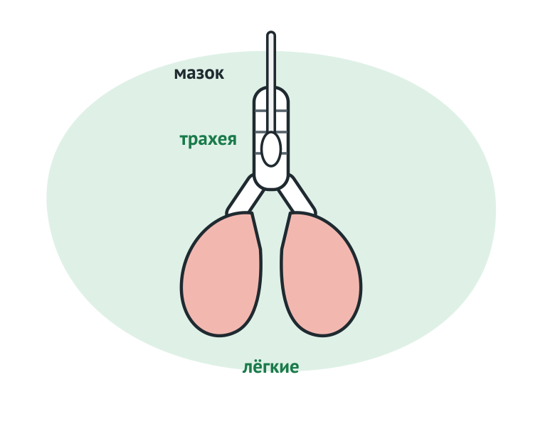
The Micoplasma colonizes the resynthetic epithelial trachea and bronchus, and the material is taken closer to the infection site. "Best" does not mean "sensitive". According to the data cited by Oh and co-authors, at an early date the larynx swabs studied by PCR are higher than both nose swabs and tracheobronchial lava. Suckers will have their larynx swabs used to assess whether a sow's agent is being transferred.
To oversee, the tracheal samples are combined into pools. According to the 2024 estimates, PCR sensitivity was 0.96 for a pool of three samples, 0.95 for a pool of five and 0.93 for a pool of ten. Pulling reduces the cost of research, and it remains the main constraint on oversight programmes.
The serology complements the PCR, but answers another question as to whether the group met with the agent.
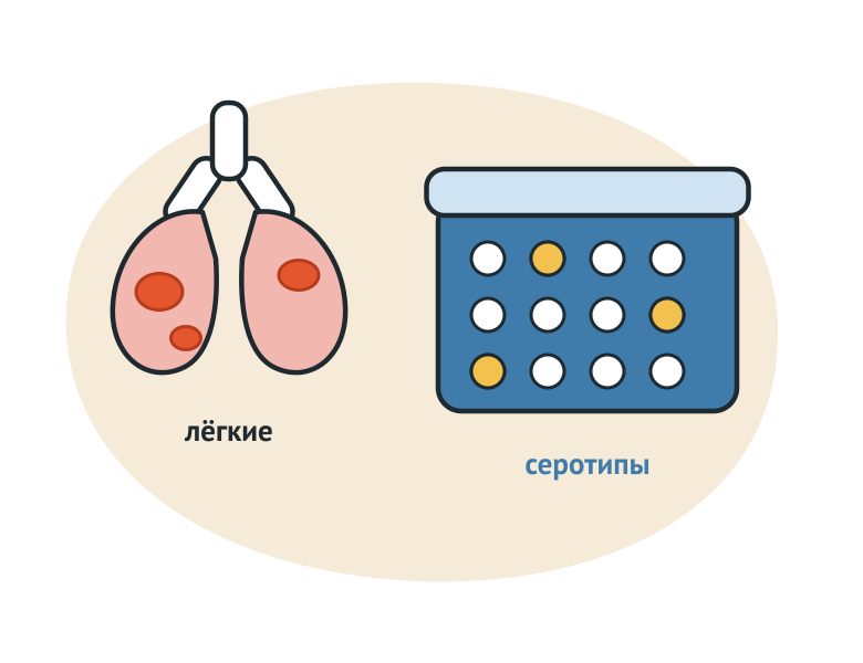
Material depends on who's being investigated. The fallen animals are taken from the affected lung tissue, almonds, bronchial and amphibian lymph nodes. The living are swabbed with mucous mouths and amydalins. By recruitment description PCR-APP-FAKTOR, a healthy animal bacterion-drive agent is separated from the almonds for 3 to 4 months after recovery.
There are 19 serotypes for surface carbohydrates, especially the capsule. Serotypes 1, 5, 9, 11 and 16 are classified as the most virulent due to toxins ApxI and AppxII. The serotype is important for the selection of a bacterial vaccine. Due to cross-sectional reactions and low reproducibility of serological methods, the serotype is determined molecularly.
The herd's serology shows another way to see if there was any contact. In an experiment with pigs infected with serotypes 1 and 7, antibodies to the lipopolisaharide were detected from the 14th to the 56th days, while the answer to ApxIV's toxin was delayed by another 14 to 21 days. This should be taken into account when choosing the time for selection of the serum.
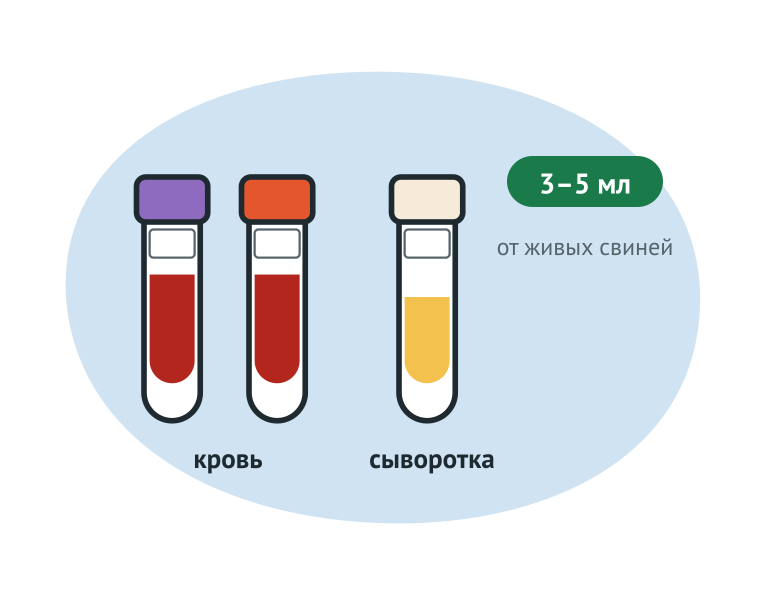
The respiratory form of the RRT is manifested by breathing, fever, denial of feed and fragrance. Young pigs are sicker; The infection often occurs sub-clinically in the cartilage and uninhabited sows.
According to the WOAH, after infection there is viremia and lung infection: young pigs have them for weeks, adults for days. So the best way to detect a virus is with serum and broncholalves. PCR applies to tissue and serum; In the case of rounding, oral fluid is suitable and in tribal herds it is appropriate to process fluids: excruciate tissue collected during castration and tail buying. Important restriction: The PCR does not distinguish the infectious virus from the activated virus.
The antibodies to the RRTS virus are detected from 7–14 days after infection, peaking at 30–50 days, and some pigs become seroneative 3–6 months later. A single serum sample is therefore not very informative: the results are influenced by maternal antibodies and past vaccinations. Maternal antibodies have a half-life of 12 to 14 days and are usually detected up to 4 to 8 weeks after birth. Seropositive sows in the infected environment are serum-converted from 3 to 6 weeks. To show a fresh infection, two samples are examined: acute serum and reconcentrated.
Suspicion procedure is set Veterinary rules for RFMOsapproved by Order No. 625 of the Ministry of Agriculture of Russia of 26 October 2020. They are in operation until 1 September 2032.
Diagnosis is considered to be established if the agent has been identified, its genetic material has been detected or antibodies have been found that are not related to vaccination (p. 18). The latter clause determines the value of vaccination information in an accompanying letter.
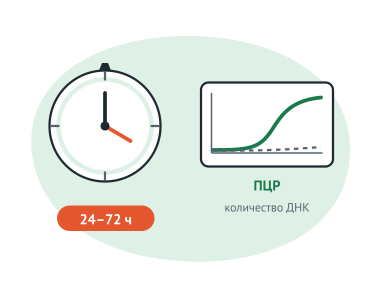
У гриппа окно для ПЦР короткое. Признаки и выделение вируса с носовой слизью начинаются уже через сутки после заражения, а выделение обычно прекращается к 7–10-м суткам. Руководство ВОЗЖ рекомендует отбирать пробы в течение 24–72 часов после появления признаков и брать материал от нелеченого животного в острой фазе. РНК ищут в трахее, лёгких и носовых мазках. Пробы везут охлаждёнными на льду, не замораживая. Заболеть при гриппе способно всё поголовье группы, смертность обычно низкая, но вторичные бактерии утяжеляют картину.
The flu serology answers the question about past contact. Historically, the main test was RTGA on steam serums taken from 10 to 21 days apart; The rise of the titre four times and more indicates a recent infection.
With CBS-2, the other is complex: the virus is widespread, and the fact that it's discovered means little. The value is the number. In experimental work, high viremia was regarded as 7 log10 copies of the genome per millilitre of serum and above. System disease was confirmed by a combination of three conditions. One: loss of mass. Second: Moderate or expressed lymphoid depletion with granulematose inflammation of lymphoid tissue. Third: Moderate or high amount of nucleic acid CBS-2 in these hybridized in situ tissues. Therefore, positive PCRs in CEF-2 are read together with clinical painting and pathomorphology.
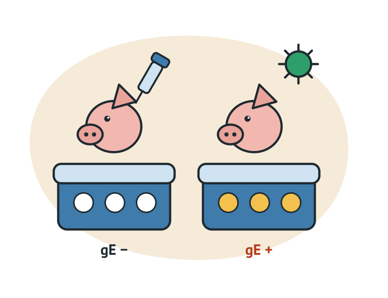
Свиньи, перенёсшие болезнь Ауески, остаются латентно инфицированными; исключение составляют поросята младше двух недель, которые гибнут от энцефалита. Латентных и субклинически заражённых животных выявляют серологией, а не выделением возбудителя. Антитела появляются на 7–10-е сутки после заражения.
Marked vaccines are free of one glycoroteine, most often gE. The inoculated non-infected animal does not give antibodies to gE, but the infected one does. This is the basis for the DIVA principle. The level of punctual immunity is assessed by other tests: gB-ELISA, latex agglutination or neutralization of the virus. A real-time PCR has also been developed, distinguishing gE-deployed vaccine virus from the wild genes gB and gE.
Veterinary rules for the illness of Aureskiapproved by Order No. 540 of the Ministry of Rural Affairs of Russia on 14 September 2020, is valid until 1 March 2027. Living animals take flushes from mouth and nose, 5 ml of blood with anticoagulant, and 5 ml without it. 18). The corpses are taken from fragments of parenchimatose organs and (or) brains in 5–10 g, and (or) lymph nodes in their entirety, and (or) almonds (p. 18). Samples shall be delivered to the laboratory within 12 hours of selection (p. 19).
Diagnosis is established in one of the five cases (p. 21). An agent has been identified or its genetic material has been discovered that is not related to vaccine strains. The results of the biotest are positive. They found antibodies that aren't vaccine-related. Finally, antibodies to glycorothein gE were found in animals who had been vaccinated with a marked vaccine. The latter case is important for practice: the positive gE-ELISA in the inoculated herd is a basis for diagnosis rather than a punctual trail.
Pasteurella multitocida and Bordetella bronchiptica are more likely to be the second layer, with already damaged mucus. The PCR finds them fast, but there's not enough PCR to diagnose.
Veterinary rules for pasteurella, утверждённые приказом Минсельхоза России от 31.10.2022 № 770, действуют с 1 марта 2023 года до 1 марта 2029 года. Диагноз считают установленным, если возбудитель выделен и идентифицирован (п. 21). Методы: бактериологическое исследование либо ПЦР с определением патогенности возбудителя биологической пробой (п. 20). От животных берут 2–5 мл крови и слизь из носовой полости; патматериал отбирают, если с момента гибели прошло не более 5 часов (п. 18). Пробы доставляют в течение 24 часов (п. 19).
The herd's serology is read only with the vaccination scheme. Four practical rules:
Inoculation information is not a formality: Regulation No. 625 refers to it in the accompanying letter for samples (para. 17) and the lab relies on them to interpret the result.
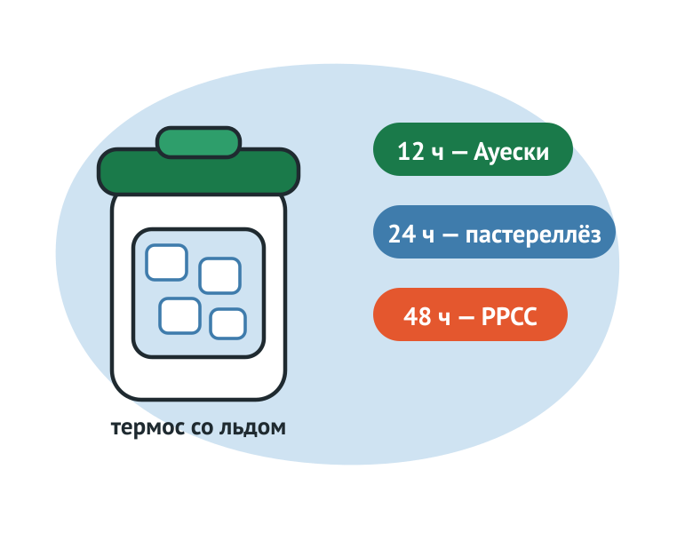
Delivery times vary by disease, and this tends to fail.
| Disease (document) | Main samples in suspicion | Date of delivery to the laboratory |
|---|---|---|
| RPTC (Order No. 625) | Blood from the living 3 - 5 ml; spleen and lungs 5 - 10 g, exhumed chest cavity, amplified lymph nodes from corpses | 48 hours (p. 17) |
| Aueski disease (Order No. 540) | Washing from mouth and nose, blood 5 ml with anticoagulant and 5 ml without it | 12 hours (p. 19) |
| Pasteurelles (Order No. 770) | Blood 2 - 5 ml, mucus from the nasal cavity; Painting takes no later than 5 hours after death | 24 hours 19) |
The general requirements are the same: the samples shall be cooled, the containers sealed in ice or chiller thermos for the duration of carriage. In order to release the RRT virus, the WWS management recommends that the material be cooled to 4°C immediately after selection and delivered within 24 - 48 hours.
PCR in real time:
ELISA ID.vet:
For nucleic acid extraction before PCR: DNA/RNA-S-FAKTOR (cat. № EDR001-VET).
VET FAKTOR specialists can help select kits for your laboratory’s needs and equipment: +7 (495) 789-48-11, vet@faktor.ru.
The agent colonizes the resynthetic epithelial trachea and bronchus, so he takes breather samples. According to the data cited by Oh and co-authors, early exposure to lubricant is higher than that of PCR and nose swabs, and tracheobronchial lava.
For tracheal samples on M. Hyopneumoniae's bullet was studied. The calculated response was 0.96 for a pool of three samples, 0.95 for a pool of five, 0.93 for a pool of ten. Bullets reduce the cost of supervision, although sensitivity is a little lower.
Not if a marked vaccine without gE was used. Cf. 21 of Regulation No. 540 detection of antibodies to glycoroteine gE in animals infected with a marked vaccine is one of the reasons for considering that the diagnosis of Auresca's disease has been established.
According to the WOAH, samples are taken within 24 - 72 hours of the appearance of signs from an untreated animal in acute phase. The virus is usually stopped by the 7th to 10th days, and later PCR may be negative if the infection is actually carried out.
Nope. Cf. 21. Regulation No. 770 is considered to have been established if the driver has been isolated and identified. PCR under art. 20 are applied together with the determination of the pathogenicity of the agent by biological sample.
This material is for reference only and does not replace veterinary rules or a veterinary professional’s assessment. Diagnosis is established on the basis of laboratory findings in accordance with the procedure prescribed by law.


© 2015–2026 VET FAKTOR LLC. All rights reserved.