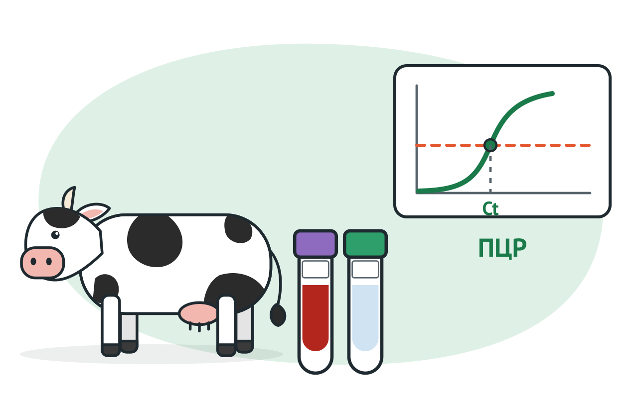
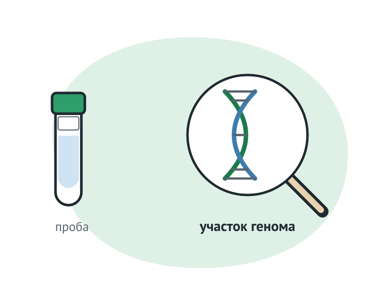
The PCR duplicates the target area of the driver's genome on multiple occasions. According to the WOAH, the most common methods of detecting nucleic acids are PCRs with its variants and insulated amplification. Reactions are seen in different ways: agarose gel, sword probes (e.g. TaqMan), real-time accumulation of the product. FACTOR PCR sets are designed for real-time PCRs, and there is also an option for the EPS virus with electrophoric separation.
Molecular methods detect genomic sequences of both living and dead microorganisms as long as nucleic acid remains in the sample. Hence the frequent discrepancy: the sample is positive in PCR and negative when the virus is released. The WOAH recommends that such cases be dealt with by sequencing or other molecular method.
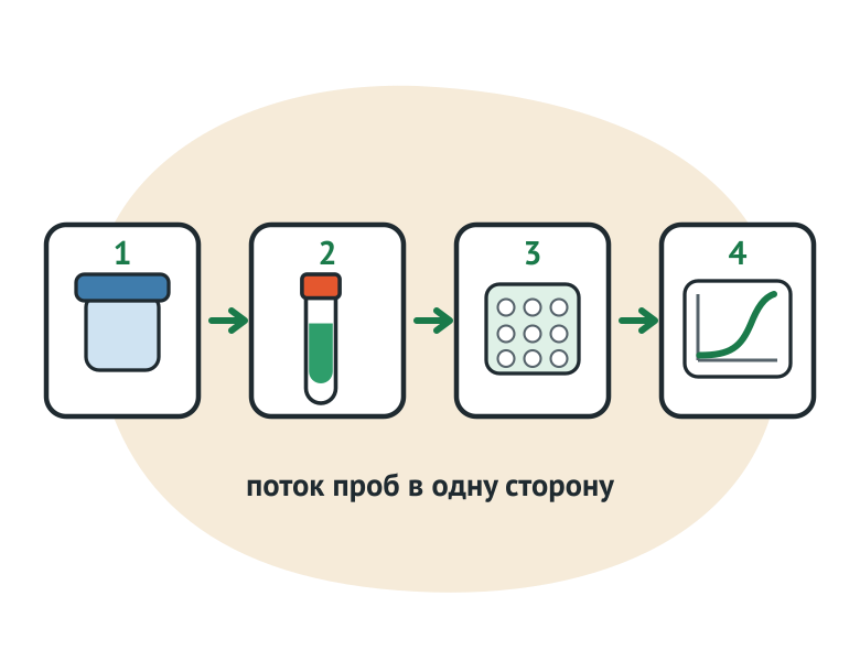
The analysis breaks down to four steps: reception and primary treatment of the material, nucleic acid emission, reaction mixture preparation and amplification, accounting for the result. MC 1.3.2569-09 converts these steps into consecutive working areas (p. 5.8):
Reactional mixtures shall be prepared prior to the delivery of samples from zone 2 to zone 3 (para. 5.11.1). Zone 3 itself is recommended to be divided into sub-zones 3a (mixtures and reverse transcription) and 3b (implicity and counting) in separate premises (p. 5.11.2). Zones 4-1 and 4-2 are placed in isolation from zones 1 to 3 so that the amplification products do not enter with an air stream; They are prohibited from being united (p. 5.12, 5.12.3). The furniture and equipment of each zone shall be marked, not used in other areas (para. 5.14.2). In zone 3, air flow is equal to extraction, in zones 1 and 2 the extraction takes precedence (p. 5.13.1, 5.13.2). The measuring instruments shall be verified at least once a year (p. 5.15).
The leadership of the WOAH requires the same in meaning. Due to the high sensitivity of the method, separate rooms or boxes are needed for individual stages, change of robes and gloves, and a strict cleaning programme.
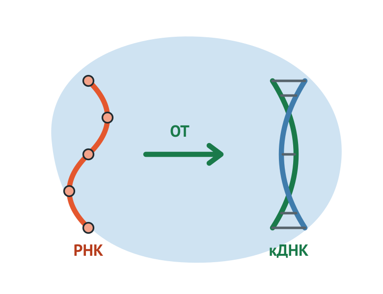
The selection begins with what is made up of the driver's genome. DNA targets are, for example, the African swine fever virus, the noduular dermatitis virus, and the CDS leukemia virus. RNA target, for example, influenza A virus.
For RNA, the reverse transcription is used before amplification: the enzyme synthesizes the CDN and then goes on to a normal PCR. That's why the set PCR-INFLUENZA-A-FAKTOR It works in the PCR format. The DNA/RNA-C-FACTOR manufacturer's description kit is designed for DNA/RNA extraction followed by reverse transcription and PCR.
Three practical conclusions follow from this. In the control list for the RNA test, the WWR adds the control of reverse transcription. The MIQE manual requires the evaluation of residual genomic DNA in RNA drugs: for this purpose, Cq is compared with and without reverse transcription. RNA viruss are changing rapidly, sharp mutations are possible, so the WWD recommends periodically checking the sequences of primers and probes.
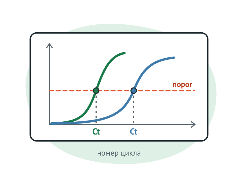
The instrument measures fluorescence after each cycle. The cycle number at which the signal crosses the threshold is given in the Ct (cycle threshold) manual by the MIQE - Cq (Quantification cycle).
Cq shows the position of the amplification curve relative to the axis of the cycles and is directly related to the target reference concentration: the more copies in reaction, the earlier the curve crosses the threshold. The restrictions begin. The values of Cq differ between plants and instruments, so they cannot be compared between laboratories. They depend heavily on the effectiveness of PCRs, which vary from set to set and even from sample to sample. The variability is influenced by threshold setting, response efficiency, quantity of source material, reaction artifacts, pipetration errors and sample variation.
The cut-off threshold sets the instructions for the set. The WWHA gives an example: sometimes the threshold is about 35 cycles, and then large Ct samples are classified as negative or questionable. For another PCR, any registered Ct is considered positive. The values between the positive and negative thresholds are called a questionable zone. In choosing the threshold, account is taken of the characteristics of a particular PCR, validation data, purpose of the result and animal veterinary information.
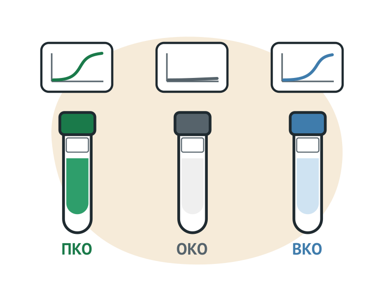
The CDS noduular dermatitis and leukemia are listed in the AFS, influenza A kits cards: 55 or 110 reactions, including controls (COE, PCO, COE). The decryption and the accounting rules are looked at in the instructions for the set. They cover different risks in terms of what they mean.
| Monitoring | What Shows | How often they do. |
|---|---|---|
| Negative sample without matrix | Contamination: signal where the product must not be | About 5% of the holes, randomly on a tablet. MIQE: In each production, download the last |
| Positive | That the reaction has passed; its Ct falls into the specified operating range | for each tablet |
| Internal, inhibitors | That there is no substance in the sample that suppresses reaction. | for each sample if inhibitors are essential to this matrix |
| The host species. | That the sample actually came from the right species of animal. | for "indirect" samples such as those collected on tampon, to each sample |
| Reverse transcription | I'm sure it's been a fusion phase of the KDNC. | in RNA tests |
Positive control in the form of plasma checks amplification, but not the quality of discharge. A well-known positive sample is needed to evaluate the selection. Internal control didn't work, which means there are inhibitors in the sample, and the negative result on the target is unsatisfied. MIQE emphasizes separately: positive controls are needed where there is little or no confidence in the negative outcome, i.e. it is when the pathogens are detected.
The stability of the set is monitored by reference maps of Ct values for working test samples and through participation in inter-laboratory comparative tests.
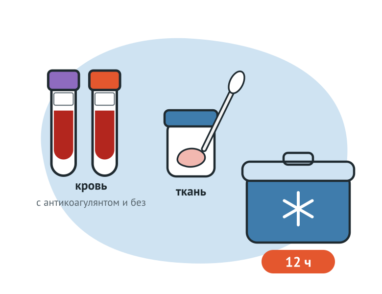
The results of the laboratory depend on the quality and suitability of the samples. Material is defined in the assignment of each set.
| Set (cat. №) | Target | Recruitment card material |
|---|---|---|
| PCR-ASF-FAKTOR (D10415-VET) | DNA of the AFP virus | Whole blood, plasma and serum, swabs with mucous nos throat and almonds, fragments of tonsils, spleen, lungs, liver, lymph nodes, etc. |
| PCR-BOVINE-LUMPY-SKIN-DISEASE-FAKTOR (D21416-VET) | DNA of noduular dermatitis virus | Splattered skin coverings, organs, whole blood, mucus and rot throat swabs, milk, sperm |
| PCR-INFLUENZA-A-FAKTOR (R10515-VET) | RNA influenza A virus | feces and diarrhea, swabs with mucous throat and trachea, bird cloaks, internal organ fragments, chicken embryos, feed and animal food |
| PCR-BOVINE-LEUKOSIS-FAKTOR (D11816-VET) | DNA of the CDS leukemia virus | Whole blood; Food (farsh, sausage, spy, etc.) |
Part of the matrix is known to be difficult. Fecalia, sperm and autolysed tissue usually contain more inhibitors than blood and culture. The cloak samples give inhibitors more often than the oral sample. The tampon material is also influential: some tampons contain substances that suppress reactions. Individual tests are incompatible with heparin, formalin, dry ice and even freezing.
Time limits and anti-coagulants are set by the veterinarian rules for a particular disease:
Samples on tampons shall be kept wet in the transport environment recommended for a particular test. Fecalia are kept chilled, for example at 4 °C or on ice, and examined as soon as possible after selection.
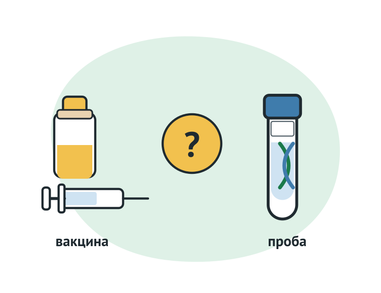
The positive result is one thing: there's a target in the sample. Further details are needed.
The legal weight of the result is determined by the rules for a particular disease. Diagnosis on the AES is considered to have been established when the agent and/or detection of its antigen, and (or) genetic material, and (or) an antibodies to the agent (para. 28 of Regulation No. 37).
In Regulation No. 156 on leukemia, the list of reasons for diagnosis is different: hematological, histologological and serological studies. The PCR is not listed in this list, although molecular-biological studies require regulations to be carried out using the PCR method (para. 20, 21). Here and above, Regulation No. 156 is cited in the wording in force since 1 September 2025: Order of 8 April 2025 by the Ministry of Agriculture of Russia. No. 229 amended paragraphs 18, 19 and 21 and supplemented the rules with paragraph 21 (1).
The laboratory part of the review is based on the guidance of the OWG:
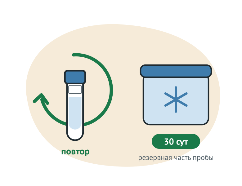
The two duties of the laboratory are given by Regulation No. 37 whether or not the result is questionable. All samples delivered are divided into two equal parts. The second shall be stored for at least 30 calendar days at a temperature between minus 15 and minus 40 °C; it shall be re-examined upon request by those contesting the result (p. 26). The head of the laboratory shall report the results in writing within 12 hours to the head of the regional veterinary authority and to the public service specialist who sent the material. 29).
S.P.H. 3.3686-21 (Decision of the Chief Public Health Officer of the Russian Federation of 28 January 2021 No. 4), in para. 140 refers to the operating requirements of laboratories using nucleic acid amplification techniques when dealing with material containing micro-organisms of groups I to IV pathogenicity. The specific document does not refer to the paragraph. The current document with this title is MM 1.3.2569-09, approved on December 22, 2009: Zoning, sample accuracy, equipment marking, ventilation, instrument verification.
The management of the SWD adds a quality system. A quality management system is needed to recognize the results and accreditation on ISO/IEC 17025. It includes validated methodologies and proven expertise, including through regular participation in inter-laboratory comparative tests.
Specific samples, timing and reasons for diagnosis are sought in the veterinarian sickness regulations. A comparison between Regulation No. 37 and Regulation No. 156 shows how different the role of PCR is for different diseases.
B Section PCR A catalogue of over 130 sets of in-house production, more than 100 of them for animal disease, bird, fish and bees. It's on the recruitment card, most of them have 55 and 110 reactions.
To extract nucleic acids:
Examples of PCRs for different types of targets:
Instructions are opened in the personal office upon confirmation of access and may also be requested by the "Ask Instruction" button on the recruitment card. VET FAKTOR specialists can help select kits for your laboratory’s needs and equipment: +7 (495) 789-48-11, vet@faktor.ru.
PCR's looking for nucleic acid as an agent, ELISA is an antibodies or antigen. Regulation No. 37 allows both methods (para. 27). Diagnosis on the AES is considered to have been established when the agent and/or detection of its antigen, and (or) genetic material, and (or) antibodies to the agent (p. 28).
Nope. Ct relates to the number of copies of the target in response, but depends on the threshold, the effectiveness of the PCR, the quality of the discharge and the instrument. The values are not comparable between laboratories, so they are not transferred from another's protocol or treated as a measure of gravity.
This discrepancy is described in the WOAH manual: molecular methods identify the sequences of both living and dead microorganisms. Discouraging by sequencing or other molecular method shall be taken.
The reasons are that internal controls (indicators in the matrix) have failed, delivery times have been violated, and material is not suitable for testing. All three make the negative result uninformative.
Research into the detection of nucleic acids from PBA is done on the requirements for laboratories using amplification techniques (p. 140 San Pin 3.3686-21). MC 1.3.2569-09 allows not only separate rooms but also the selected working areas of other premises, while maintaining a confinity (para. 5.2, 5.8).
This material is for reference only and does not replace veterinary rules or a veterinary professional’s assessment. Diagnosis is established on the basis of laboratory findings in accordance with the procedure prescribed by law.


© 2015–2026 VET FAKTOR LLC. All rights reserved.