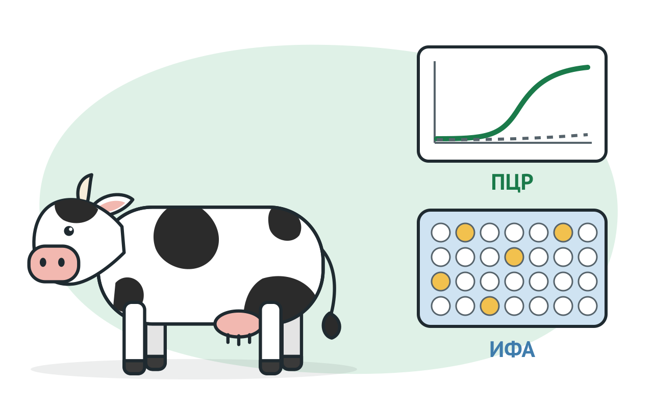
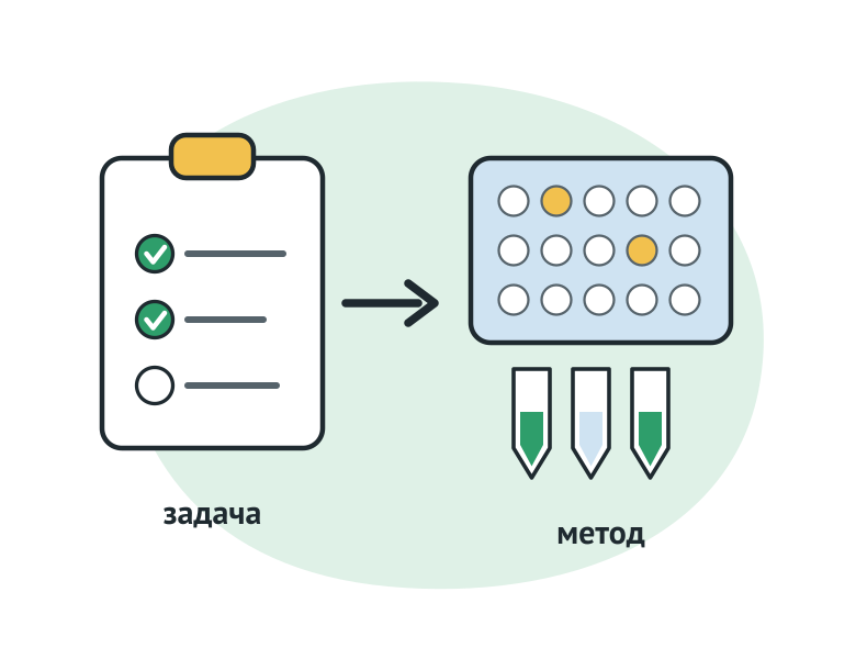
The WOAH Manual on Diagnostic Tests (Terrestrial Manual, chap. 1.1.6. Calls for the six most frequent laboratory tests:
The kit is validated for destination, species of animals and type of sample. This applies to both laboratory methods and animal expresses.
Different goals require different test properties. The screening is applied to a healthy population, so it's expected to be highly diagnostically sensitive: missing an infected animal is more expensive. The validation test solves the reverse task of reducing the percentage of false positive results. Hence the frequent pattern: sensitive screening, then specific confirmation. In assessing the punctual immunity, a false positive result is particularly dangerous: an animal is considered protected, although it does not have immunity.
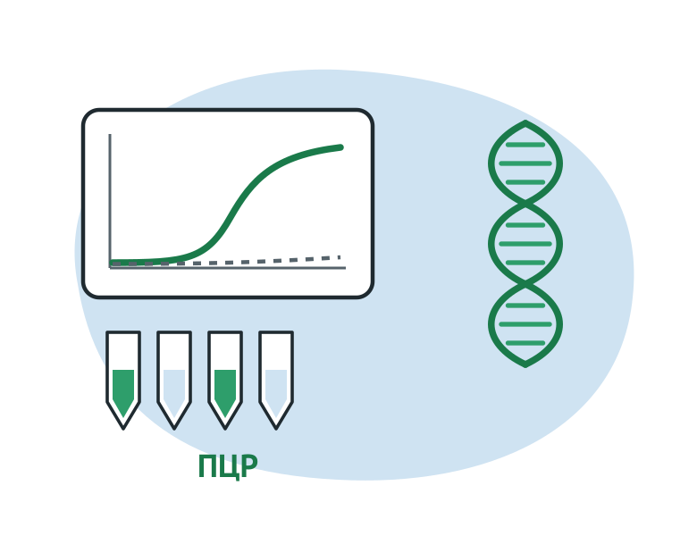
The PCR identifies a part of the driver's genome. It's a direct method: it answers the question whether there is genetic material in the sample. That's why it's important. The PCR finds sequences of both living and dead microorganisms as long as nucleic acid remains in the sample. The crop needs a living booster to grow.
Before a reaction, nucleic acid is extracted from the clinical material by a separate set. The WOAH recommends that the extraction method be chosen in advance: with large sample streams, automatic extraction may be necessary.
High analytical sensitivity to amplification also creates risk. The transfer of amplions from sample to sample gives false positive results. The WWS recommends separating the steps by room or box, changing the robes and gloves, and maintaining cleaning programmes.
The result is read with the controls:
The inhibitors are more common in feces and sperm, less frequent in blood. The cloak samples contain them more often than those of the mouth.
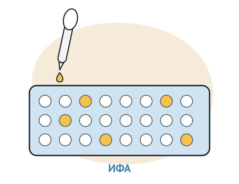
ELISA detects antibodies or antigens in the serum, blood plasma and other samples. The method is used for serological monitoring of the population, screening and assessment of immunity after vaccination.
Formats differ by what's on the tablet and what they catch:
| Format | How it works | Where you're comfortable |
|---|---|---|
| Indirect | adjacent antibodies of the sample are detected by an anti-species conjugate | скрининг больших количеств проб; один набор рассчитан на один или несколько видов животных |
| Competing (locking) | The antibodies of the sample compete with the sword antibodies for the antigen on the tablet. | Species for which no anti-species conjugates exist; The serum is taken in low breeding, less interference with non-specific binding |
| Sandwich | Antigen is holding a sorbine antibody trap. | Large antigens and complex proteins; Limiting format - the antigen must have at least two antigen plots. |
ELISA for antigens and PCRs solves the common problem of finding the very agent. Antigen tests and a diagnostic window are usually more visible than antibody tests: the immune response holds longer than the driver in the test.
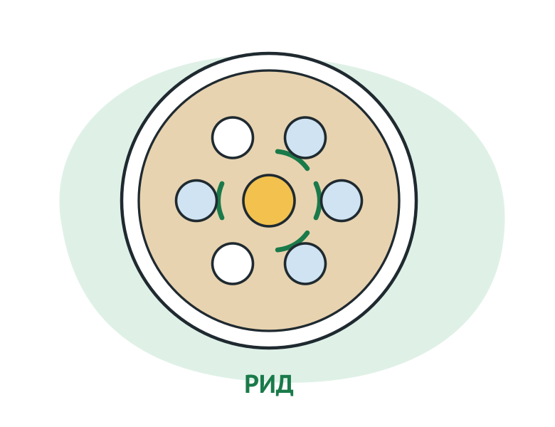
RID is an immunodiffuse reaction in agaric gel (AGID). The serum and the antigen are used in adjacent gel holes. The meeting will produce a line of precipitation and be read in the eye. The equipment's simple, it's not expensive.
The RWD considers RID to be less common antibodies detection techniques. For leukemis, the CRS management describes RID as a specific but not very sensitive test for seruming individual animals. It's not good for milk except the first molesus.
Practical conclusion: RIDs are well-confirmed and weaker. When a herd's serology requires sensitivity, ELISA or PCR are added to it.
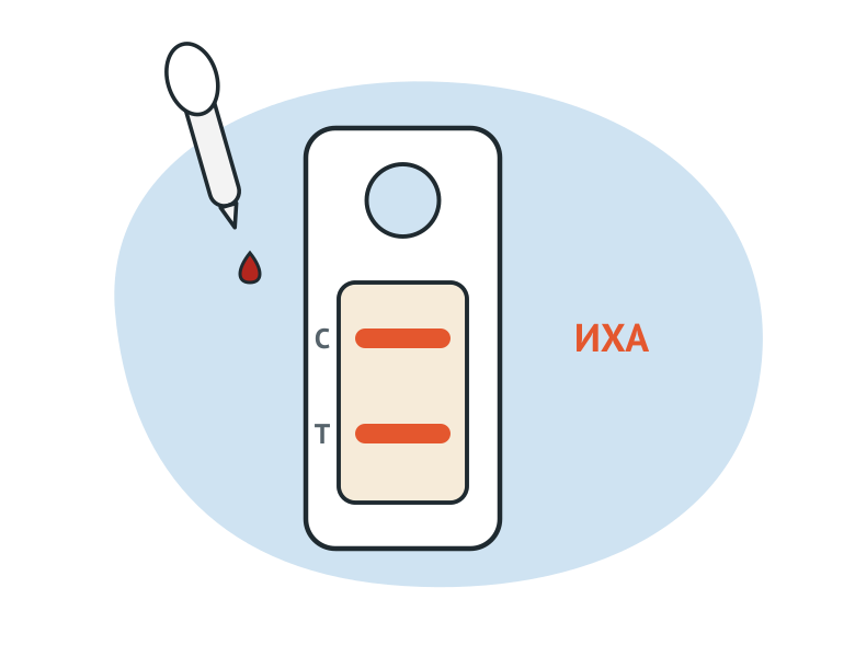
Immunochromatographic analysis (ICA) is done on a test field or in a cassette. WWSS calls such devices point of care, or pen-side tests, and assigns them to new antibodies detection techniques.
The conditions of work of the WWHA are clearly described. Express tests are applied in the field under different ambient conditions, on different types of samples collected under non-sterile conditions by operators with different training. The test is therefore required to be resistant to temperature, humidity and illumination fluctuations and to the operator's qualifications. OSW points out that the need for evidence-based research and reporting by an accredited positive results laboratory – especially in the study of exotic diseases – should be enshrined in existing testing rules and guidelines.
Hence the role of IHA: quickly select animals for further research, get a preliminary assessment where the lab is far away. Diagnosis is determined from laboratory studies in a manner that sets veterinary rules.
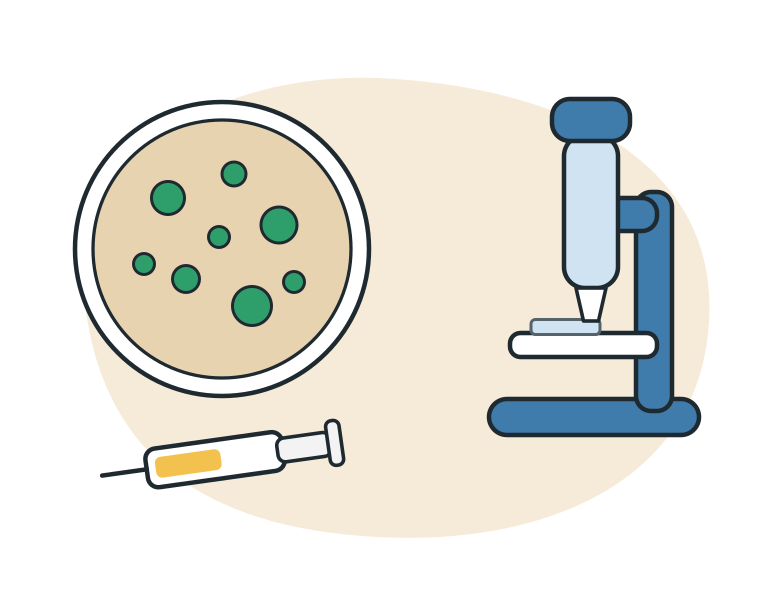
Classic methods remain in the rules where a living agent or reaction of cellular immunity is needed.
Bacteriologic research on Russian vets includes bacteria, a cultural method with a pure culture of the driver and a biological sample. The crop needs a living agent, which is different from the PCR. For Regulation No. 534 the PCR for tuberculosis is used as an express test, but the result is necessarily confirmed by a bacteriological study (para. 25).
The allergic sample is not the same: a drug for diagnosis of tuberculosis is injected into the animal and the reaction is taken into account. The planned allergic studies are being conducted to prove that there is no circulation of tuberculosis in the household (p. 8 Regulation No. 534).
Histologic research is looking for characteristic changes in the tissue. Regulation No. 156 also considers leukemia diagnosis to be established when such changes are detected in a histological study (para. 21).
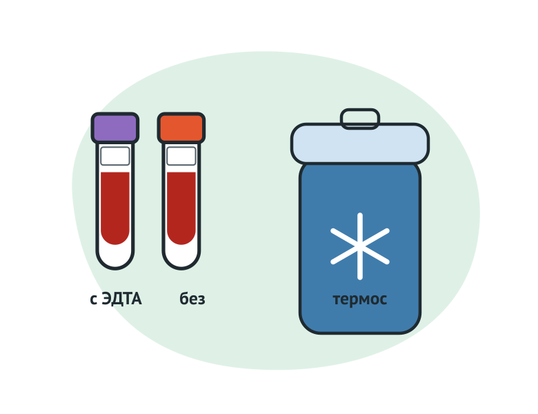
Chapter 1.1.2 of the WOAH manual starts with a simple saying: the study depends on the quality and suitability of the samples. Four frequent casualties:
The serum is derived from blood without anticoagulant: the clot is reduced at room temperature, protecting it from heat and cold. The feces samples are kept chilled – in the fridge at 4 °C or on ice – and examined as quickly as possible.
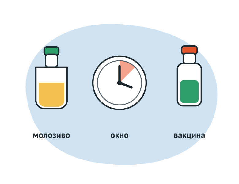
The young man's epic antibodies. The calf gets an antibodies with a mole. For the CRS's leukemia, the OSW management points out that maternal antibodies can disappear for up to 6 to 7 months and cannot distinguish them from the antibodies of active serology. Active infection confirms the PCR on the virus. Carrots with braid antibodies are listed in the manual in cases where PCR for an individual animal is useful. Regulation No. 156 made this the norm: animals from 15 calendar days to 6 months inclusive are examined by molecular-biological methods.
The window's up to the seroconversion. The diagnostic window is the interval between contact with the agent and the moment when the antibodies are first detected. For the CRS leukemia virus, the WWD management provides 3–16 weeks. Negative serology in a newly infected animal is expected and the disease does not rule out. The leukemia also has a long incubation period: the rules refer to 2 months to 6 years.
A case in point is the birth period. The antibodies go from cow's blood to molesic, and the RID may not see them. The WWS management does not consider the final negative result of RIDs to be 2 to 6 weeks before departure and 1-2 weeks after; The study is repeated. Regulation No. 156 determines this time limit: The blood is taken for serology at least 14 calendar days after the removal and no later than 14 days before it.
Vaccination antibodies. Serology shows contact with the antigen, and its source is vaccine. Regulation No. 703 requires that the date of last vaccination, the series number, name and manufacturer of the vaccine (para. 23). These data are provided if they are submitted by an animal owner or a veterinary specialist. When there was no vaccination, the letter says it is. Regulation No. 156 does not permit the taking of blood for serology until 14 calendar days after the introduction of living vaccines or allergens (para. 18). If the vaccination data didn't reach the lab, the results would be misread.
| Method | What it detects | Sample | When used |
|---|---|---|---|
| Real-time PCR | DNA or RNA of the driver, including the deceased | On instructions for recruitment; for the CRS's leukemia virus – whole blood and food | Confirmation of the case; Young people with porcine antibodies; period to seroconversy |
| ELISA on antibodies. | Antibodies to the Inducer | serum or plasma, in part of the sets - combined samples | Head monitoring, screening, post-vaccination immunity assessment |
| ELISA on antigen. | Antigen of the booster | on instructions for recruitment | The period during which the agent is released; The window is more than serology. |
| AGID | antibodies precipitating | serum or plasma | Serology where the method is secured by rules; confirmation |
| IHA (express test) | antibodies or antigens | Test instruction | Preliminary field assessment; Positive results confirmed in the lab |
| Bacteriologic study | living agent | Paint, milk, secrets by the rules. | Diagnosis for tuberculosis and brucellosis |
| Allergic sample | Animal reaction to an injectable drug | The drug is injected into the animal. | Planning studies for tuberculosis (para. 8 Regulation No. 534) |
| Gematological study | Changes in the blood image | blood with 10% EDTA solution | CRS leukemia: in animals over 6 months after positive serology (p. 18 of Regulation No. 156) |
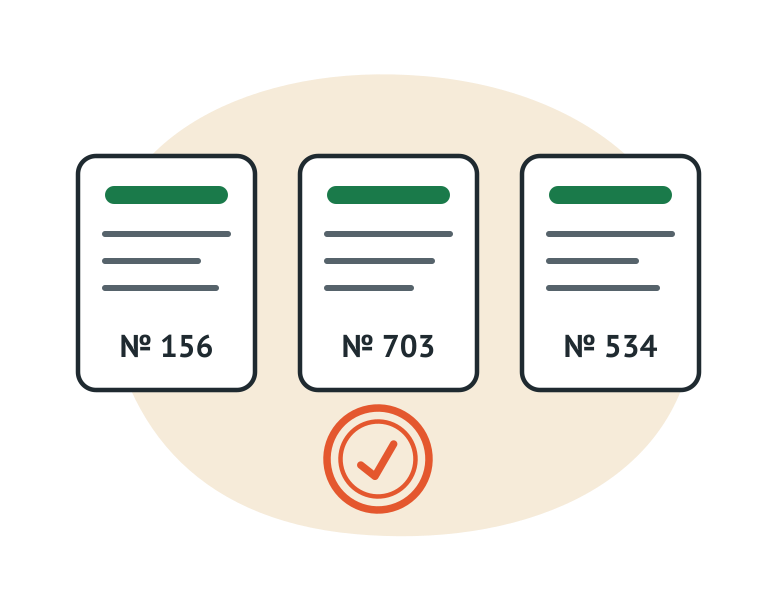
Russian veterinary rules often refer to the method directly. Three examples.
The CDS Leukose. Правила, утверждённые приказом Минсельхоза России от 24.03.2021 № 156in force from 1 September 2021 until 1 September 2027. Serological studies are conducted by ELISA and (or) RIDs, molecular biology by PCR (p. 20). The method is age-related (p. 18). Animals over 6 months take 5 to 7 ml of blood without an anticoagulant or with a twist factor, for serology. Between 15 calendar days and 6 months inclusive, animals receive 5 to 7 ml of anticoagulant blood for molecular-biological research.
Плановую серологию у животных старше 6 месяцев проводят раз в год. На быков-производителей (доноров), коров-доноров эмбрионов и животных-продуцентов это правило не распространяется: животных-продуцентов проверяют 2 раза в год, быков-производителей, используемых в случке, – 3 раза в год (п. 9). Подпункт «а» пункта 9, пункты 18, 19 и 21 действуют в редакции приказа Минсельхоза России от 08.04.2025 № 229 – с 1 сентября 2025 года. Разбор по группам животных – в статье "Leykos CRS: RID, ELISA or PCR".
Broucellosis. Rules approved by Order No. 703 of the Ministry of Agriculture of Russia of 22.11.2024in force from 1 March 2025 until 1 March 2031. Research is done by bacteriological method, PCR and serological methods from Table No. 2 of paragraph 13 of the Regulation (para. 24). When the result of serology is questionable, blood is taken again 25–45 calendar days after the day of initial selection (p. 22). Samples shall be delivered to the laboratory within 48 hours of selection (p. 23).
Tuberculosis. Rules approved by Order No. 534 of 8 September 2020 of the Ministry of Agriculture of Russia, действуют с 1 марта 2021 года до 1 марта 2027 года. Для доказательства отсутствия циркуляции возбудителя проводят плановые аллергические исследования (п. 8 в редакции приказа Минсельхоза России от 02.11.2022 № 777, действует с 1 марта 2023 года). Методы лабораторного исследования – бактериологические, включая бактериоскопический, культуральный и биологическую пробу, а также ПЦР (п. 25). ПЦР применяют для идентификации возбудителя при бактериологическом исследовании и как экспресс-тест с обязательным подтверждением бактериологическими исследованиями. Отрицательный результат ПЦР окончательным не считают. Пробы доставляют в лабораторию в течение 72 часов с момента отбора (п. 24).
The difference between methods is easy to see on one disease. For CRS leukemia, the catalogue has sets of all three methods called Regulation No. 156:
The catalog sections are arranged using the method. In section PCR – More than 130 sets of indigenous production, more than 100 of them for animal disease, bird, fish and bees. Most people have 55 and 110 reactions. In section ELISA - Over 110 sets of ID.vet for serological diagnostics and control serums of ID.vet. Separately collected IHA test field and Total Catalogue.
VET FAKTOR specialists can help select kits for your laboratory’s needs and equipment: +7 (495) 789-48-11, vet@faktor.ru.
The PCR is looking for the genetic material of the agent, ELISA is an antibodies to him or his antigen. The PCR answers the question: "The agent is here," serology: "the organ has met with him." The serology does not distinguish the transported infection from the vaccination, and the PCR identifies nucleic acid and dead microorganisms.
The private reason is colossal antibodies. According to the GWD, maternal antibodies to the CDS leukemia virus can last for up to 6 to 7 months without infection. Regulation No. 156 therefore requires molecular-biological research for animals from 15 calendar days up to and including 6 months.
Nope. The OSW indicates that the need for evidence-based research and reporting by an accredited positive results laboratory – especially in the study of exotic diseases – should be enshrined in testing regulations. Diagnosis is determined by the results of laboratory studies in accordance with the procedures established by the veterinary regulations for a particular disease.
Regulation No. 703 provides for the re-sampling of blood 25 - 45 calendar days from the date of initial sampling (p. 22). The decision to proceed is made by the public service.
In order to distinguish the punctual antibodies from the antibodies after infection. Regulation No. 703 requires that the date of the last vaccination, the series number, the name and the manufacturer of the vaccine be indicated in the accompanying letter (para. 23). This information shall be provided if submitted by an animal owner or a veterinary specialist. The lack of vaccinations is also noted. Regulation No. 156 also sets the time limit for selection: at least 14 calendar days after the introduction of living vaccines or allergens (para. 18).
This material is for reference only and does not replace veterinary rules or a veterinary professional’s assessment. Diagnosis is established on the basis of laboratory findings in accordance with the procedure prescribed by law.


© 2015–2026 VET FAKTOR LLC. All rights reserved.