Bovine leukosis is caused by bovine leukaemia virus (BLV), an oncogenic RNA virus of the genus Deltaretrovirus in the family Retroviridae. Its genome integrates into the DNA of infected cells, primarily B lymphocytes, as proviral DNA. Infection is lifelong, and antibodies to the virus persist. There is no vaccine or treatment for bovine leukosis.
The rules distinguish three stages of the disease: asymptomatic, haematological and clinical. The first two have no characteristic clinical signs. In the asymptomatic stage, antibodies to the virus are present in serum; in the haematological stage, blood lymphocyte counts remain elevated. According to the rules, the incubation period lasts from 2 months to 6 years.
According to the World Organisation for Animal Health (WOAH, WHOH), most infections are sub-clining. Persistent lymphocytosis is developed in 30-70% of infected animals, and tumors occur in 0.1-10%, usually over the age of 3. So infected animals in the herd are detected by laboratory methods.
BLV is not the only cause of tumours in cattle. The WOAH manual separately identifies sporadic bovine leukosis, encompassing lymphomas in calves and the cutaneous and thymic forms, whose cause is unknown. WOAH reserves the term “enzootic bovine leukosis” for lymphomas caused by BLV. PCR can help distinguish between them in tumour tissue: WOAH considers it useful for examining tumours, including samples collected at slaughterhouses.
Testing procedures are governed by the Veterinary Rules on Bovine Leukosis, approved by Order No. 156 of the Russian Ministry of Agriculture dated 24.03.2021. They remain in force until 1 September 2027. From 1 September 2025, the rules apply as amended by Order No. 229 of the Russian Ministry of Agriculture dated 08.04.2025.
The testing methods (paragraph 20) and the timing of blood collection in relation to vaccination and calving (paragraph 18) remain unchanged. Testing intervals, requirements for introducing animals and the procedure for establishing a diagnosis by serology have changed. The main points for laboratories are:
Paragraph 18 links the animal’s age and status to the testing method, sample and collection tube:
| Group | Method | Sample under paragraph 18 |
|---|---|---|
| Calves aged from 15 days to 6 months inclusive | PCR | 5–7 ml of blood with an anticoagulant: 3% EDTA solution at a 10:1 ratio, or sodium citrate |
| Animals older than 6 months | AGID and/or ELISA | 5–7 ml of blood without anticoagulant or with a clotting activator |
| Animals older than 6 months with positive serology | haematology | 5–7 ml of blood with a 10% EDTA solution, 0.02 ml per 1 ml of blood |
| Animals found dead | histology (paragraph 21) | 2×2×1 cm tissue samples in 10% formalin at a 1:30 ratio, within 8 hours of death or slaughter |
Serology allows ELISA and (or) RIDs to be performed, while molecular studies are conducted by PCR (p. 20). The planned serological study of livestock over 6 months is carried out once a year, and animal producers (other than the bulls used in the incident) are examined twice a year at intervals of at least 180 days (para. 9). Animals for herding are examined by the same methods within 180 days before entering the farm (p. 8).
Calves receive maternal antibodies through colostrum. According to the WOAH manual, these may persist until 6–7 months of age. Passively acquired antibodies cannot be distinguished from antibodies produced in response to infection. A seropositive calf may be uninfected, while an infected calf’s antibody levels may temporarily decline.
An Argentine study illustrates this point. Seven calves infected during their first week of life were followed. In six, antibody titres fell at 3–6 months and then rose again. Proviral loads were high in all seven by 12 months. Some calves are infected in utero: field studies summarised in a 2018 review found infection in 4–18% of newborn calves tested before colostrum intake.
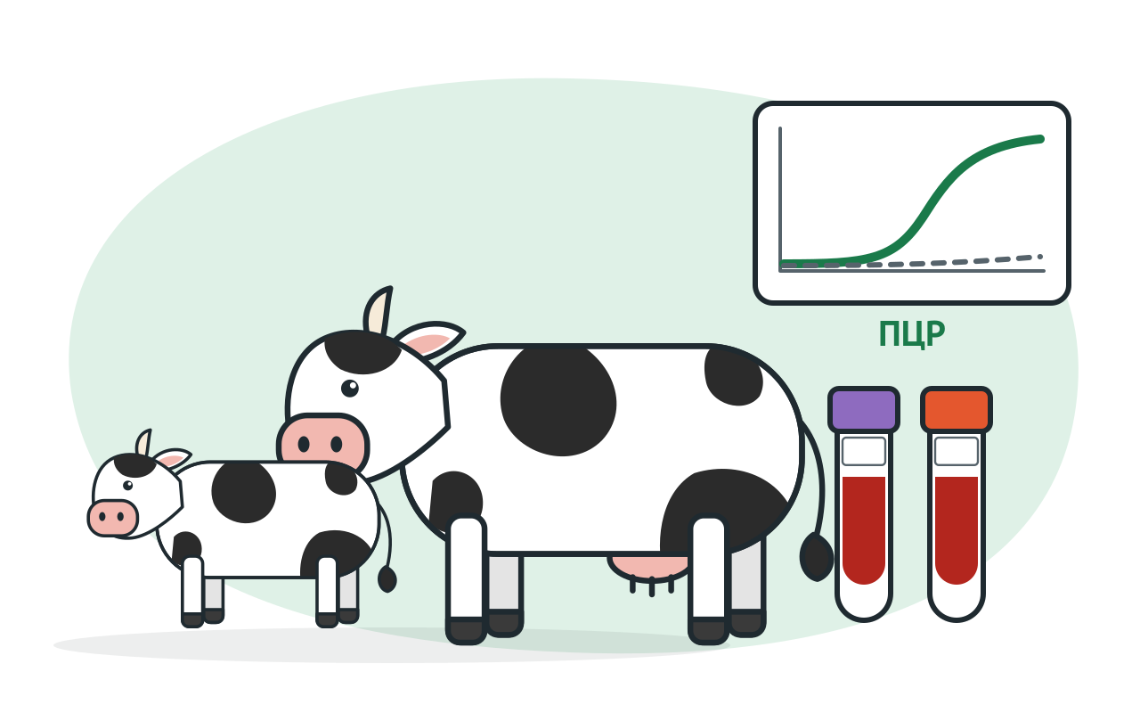
PCR detects proviral DNA and thus infection itself. This matches paragraph 18: blood with anticoagulant is collected from calves aged from 15 days to 6 months for PCR. WOAH also considers PCR useful after a weakly positive or doubtful ELISA result and following recent infection, before antibodies have appeared. Antibodies in cattle become detectable 3–16 weeks after infection.
The age of 6 months is also important for calves born from infected cows on the reservation. The only way to get them out of there is after 6 months and three negative results from serology. The interval between research is between 90 and 120 calendar days (p. 42).
A negative PCR result also has limitations. Only a small proportion of blood mononuclear cells is infected, and inhibitors in the sample can cause a false-negative result. A positive control is therefore required in every run.
Paragraph 18 sets the following timings for serological and haematological samples:
The WOAH manual describes a similar limitation for AGID. Before calving, antibodies move from the cow’s blood into colostrum. A negative serum AGID result during the 2–6 weeks before calving or the first 1–2 weeks afterwards is therefore not conclusive, and testing is repeated. During that period, AGID may be performed using colostrum from the first milking.
In order to be able to verify compliance with the deadline, an inventory of samples is attached to the accompanying letter (para. 19). It contains the sample number, animal marking device number, gender and age. It also indicates the date of the last introduction of a live vaccine or allergen and the date of departure or scheduled departure.
Under paragraph 19, samples are placed in leakproof containers, cooled and transported in an insulated flask with ice or another coolant. The containers are packaged and sealed. The covering letter states the sampling date and time, the last vaccination date, the holding, the reason for testing and the contact details of the professional who collected the samples.
The WOAH manual adds practical details:
Both methods detect antibodies. According to the WOAH manual, most tests detect antibodies to the envelope glycoprotein gp51, which appear earlier than other antibodies. The WOAH reference serum E05 is used to assess ELISA sensitivity and standardise AGID antigen.
| AGID | ELISA | |
|---|---|---|
| Sample type | Individual serum; of milk, only the first molesosy. | serum; Individual kits work with milk; Some IFAs are sufficiently sensitive to collect samples |
| Characteristics according to WOAH | specific but not highly sensitive; simple to perform | usually more sensitive than AGID |
| Result reading | after 24, 48 and 72 hours | by optical density using a microplate reader |
How to interpret AGID. A sample is positive if it forms a precipitation line with the antigen that merges with the reference serum line. A weakly positive sample bends the reference line towards the antigen well without forming a line of its own. Doubtful and weakly positive sera may be concentrated and retested. If controls produce unexpected results, the run is invalid.
How to interpret ELISA. For blocking ELISA, the WOAH manual describes an intermediate zone. An animal with a result in this zone is retested using a sample collected one month later. For some kits, WOAH advises against establishing an animal’s status from a single positive ELISA result and recommends confirmation by a second method.
The rules distinguish two statuses. An infected animal is seropositive but has negative haematology and no pathological changes. A diseased animal has positive haematology or characteristic lesions (paragraph 22). Control measures in the outbreak depend on this status: diseased animals are sent for slaughter within 15 calendar days of positive haematology (paragraph 33).
Haematology is considered positive when the age-specific thresholds per 1 µl of blood are exceeded (paragraph 22):
| Age | Leukocytes, above | Lymphocytes, absolute count | Lymphocyte proportion, above |
|---|---|---|---|
| from 6 months to 2 years | 12 000 | 11 000 | 75% |
| from 2 to 4 years | 11 000 | 10 000 | 70% |
| from 4 to 6 years | 10 000 | 9 000 | 65% |
| older than 6 years | 9 000 | 8 000 | 60% |
A diagnosis of leukosis is considered established following positive haematology or characteristic histological changes (paragraph 21). Infected animals in segregated holding facilities undergo haematological testing every 6 months (paragraph 41).
VET FAKTOR specialists can help select kits for your laboratory’s needs and equipment: +7 (495) 789-48-11, vet@faktor.ru.
The rules prescribe PCR for calves from 15 calendar days of age. They do not specify a method for younger calves.
According to the WOAH manual, there is no conclusive evidence of transmission to humans, and the virus is generally not considered a human health hazard. EFSA likewise finds no scientific basis for classifying bovine leukosis as a zoonosis.
If there are also no pathological changes, the cow is classified as infected but not diseased (paragraph 22). Within an outbreak, such an animal is isolated within 15 calendar days of antibody detection and sent for slaughter or transferred to a segregated holding facility (paragraph 33). At that facility, its blood is examined haematologically every 6 months (paragraph 41).
No. Blood for serology is collected no earlier than 14 calendar days after calving (paragraph 18). According to the WOAH manual, serum antibodies may not be detectable by AGID during the first 1–2 weeks after calving.
The rules permit both methods. According to the WOAH manual, ELISA is generally more sensitive, while AGID is simpler to perform. WOAH also notes that some ELISAs are sufficiently sensitive for pooled samples.
This material is for reference only and does not replace veterinary rules or a veterinary professional’s assessment. Diagnosis is established on the basis of laboratory findings in accordance with the procedure prescribed by law.


© 2015–2026 VET FAKTOR LLC. All rights reserved.