The disease is caused by lumpy skin disease virus (LSDV), a member of the genus Capripoxvirus in the family Poxviridae. Sheep pox and goat pox viruses belong to the same genus. The three viruses are antigenically indistinguishable, so serological tests cannot differentiate them. Cattle and buffalo are affected. The virus is not transmissible to humans.
Lumpy skin disease is listed by the World Organisation for Animal Health (WOAH) as a disease that countries must notify. In Russia, it is included in the list of contagious animal diseases (item 29.1 of the list approved by Russian Ministry of Agriculture Order No. 476). Control measures are set out in the Veterinary rules on lumpy skin disease, approved by Russian Ministry of Agriculture Order No. 588 of 24.08.2021. They remain in force until 1 March 2028.
The main losses on farms are reduced productivity: milk yield falls sharply, cows abort, and bulls may develop temporary or permanent infertility. Restrictions on cattle movement and sale during and after quarantine add to these losses.
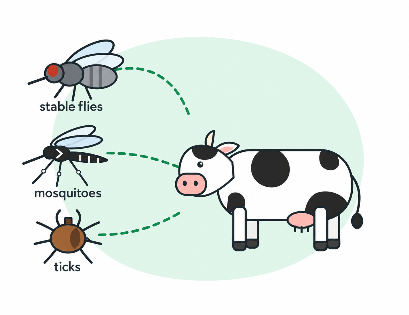
The virus is transmitted mainly by mechanical means, through blood-sucking dickstons. In their experiments, the virus was passed on to fly-girl (Stomoxis calcitrans), Aedes aegypti mosquitoes and xodium mites. The role of mites in distribution is considered small. In direct contact, without vectors, the virus is transmitted from an animal to an animal. The exception is described for the Saratov/2017 recombinant strain in Russia (for recombinants, see para. It was handed over to contact animals in an insect-protected room. In the sperm of bulls, a living virus was found for up to 42 days and its DNA was discovered for up to 159 days. In Russia, flashes were recorded from mid-May to mid-November 2015-2020.
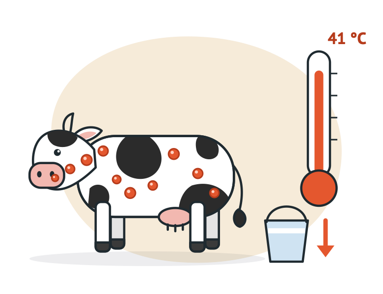
The experimental incubation period is 4–14 days; the veterinary rules specify 3 to 30 days (paragraph 4 of Rules No. 588). For international trade purposes, WOAH defines it as 28 days.
Typical presentation:
WOAH estimates that typically 10–20% of a herd becomes ill and 1–5% dies. For European breeds, EFSA reports 5–45% and less than 10%, respectively. In experiments, generalised lesions developed in only 40–50% of infected animals. The others had no generalised nodules, so suspected cases should be sampled even if obvious nodules are absent.

According to the GWD, the main differential diagnosis is pseudonodular dermatitis which causes herpesvirus of type 2 (BoHV-2). Similar skin lesions result in other diseases. In the case of mucous diseases, the list includes stork, blutang, viral diarrhoea (disease of mucus), malignant cathartic fever, infectious rinotracheitis, and papal stomatitis.
| Disease | How to investigate |
|---|---|
| Pseudo-lumpy skin disease (BoHV-2) | PCR for BoHV-2, ELISA for antibodies to BHV-2 |
| Besnoitiosis (Besnoitia besnoiti) | ELISA for antibodies to Besnoitia besnoiti |
| Dermatophilosis | Microscopy of a smear from affected skin |
| Trichophytosis (dermatophytosis) | Microscopy and culture of skin scrapings |
| Demodicosis | Microscopy of nodule contents |
| Pseudocowpox | PCR for parapoxviruses |
| Urticaria, insect bites, photosensitisation | Clinical examination and history |
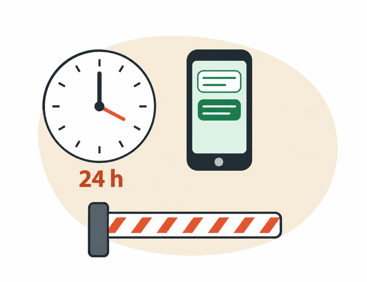
The procedure is set out in Rules No. 588:
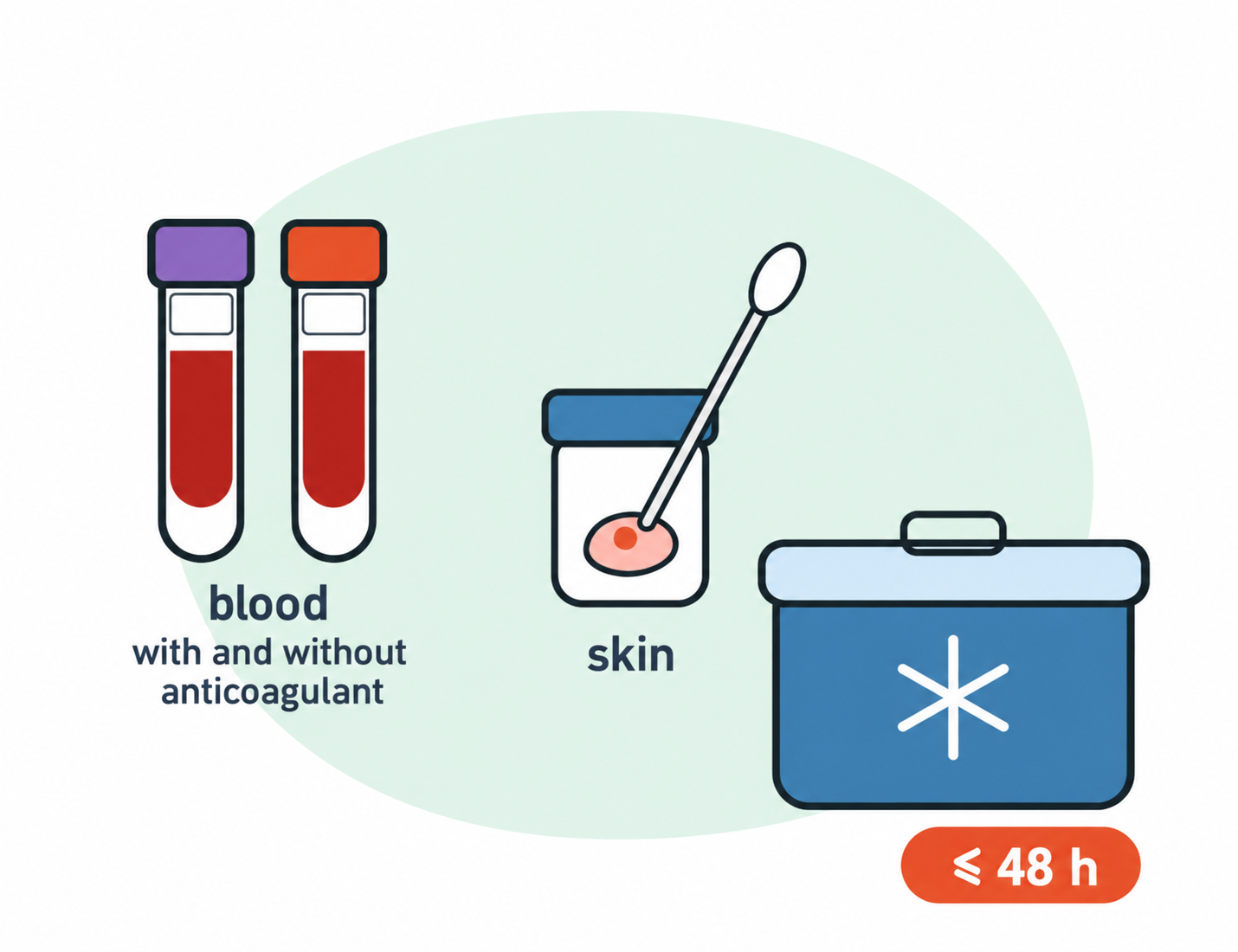
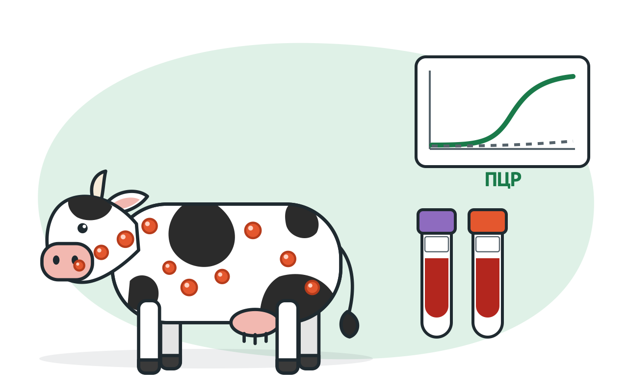
Skin nodules are the preferred samples for PCR. The viral titre in skin is five orders of magnitude higher than in blood, and DNA can be detected in lesions for up to three months. In a study of Russian samples collected in 2016, PCR was positive in 78% of skin nodule samples, 23% of nasal swabs and only 13% of blood samples.
Regulation No. 588 requires that samples be taken from each animal if in a group of up to 10 heads and 10 animals if the head is larger (para. 16).
| Sample type | When useful | Points to note |
|---|---|---|
| Skin fragments containing nodules and scrapings of affected nasal mucosa, 5–10 g | Throughout the clinical phase | Main sample for confirmation |
| Blood, 5 ml, with and without anticoagulant | During viraemia: from day 4 to day 21 after infection, depending on the study | A negative blood PCR result does not rule out the disease |
| Swabs of the conjunctival and nasal mucosa | When clinical signs are present | Complement skin samples |
| From dead animals: nodular skin, lungs, trachea and whole lymph nodes | At necropsy | Collect enlarged, oedematous lymph nodes |
Samples are placed in leak-proof containers, chilled and transported on ice in an insulated container. They must reach the laboratory within 48 hours of collection (paragraph 17). WOAH recommends that blood should not be frozen. The accompanying documentation must specify the date of the last vaccination, the vaccine batch and manufacturer, or state that the animals were not vaccinated (paragraph 17). The laboratory needs this information to interpret a positive PCR result correctly.
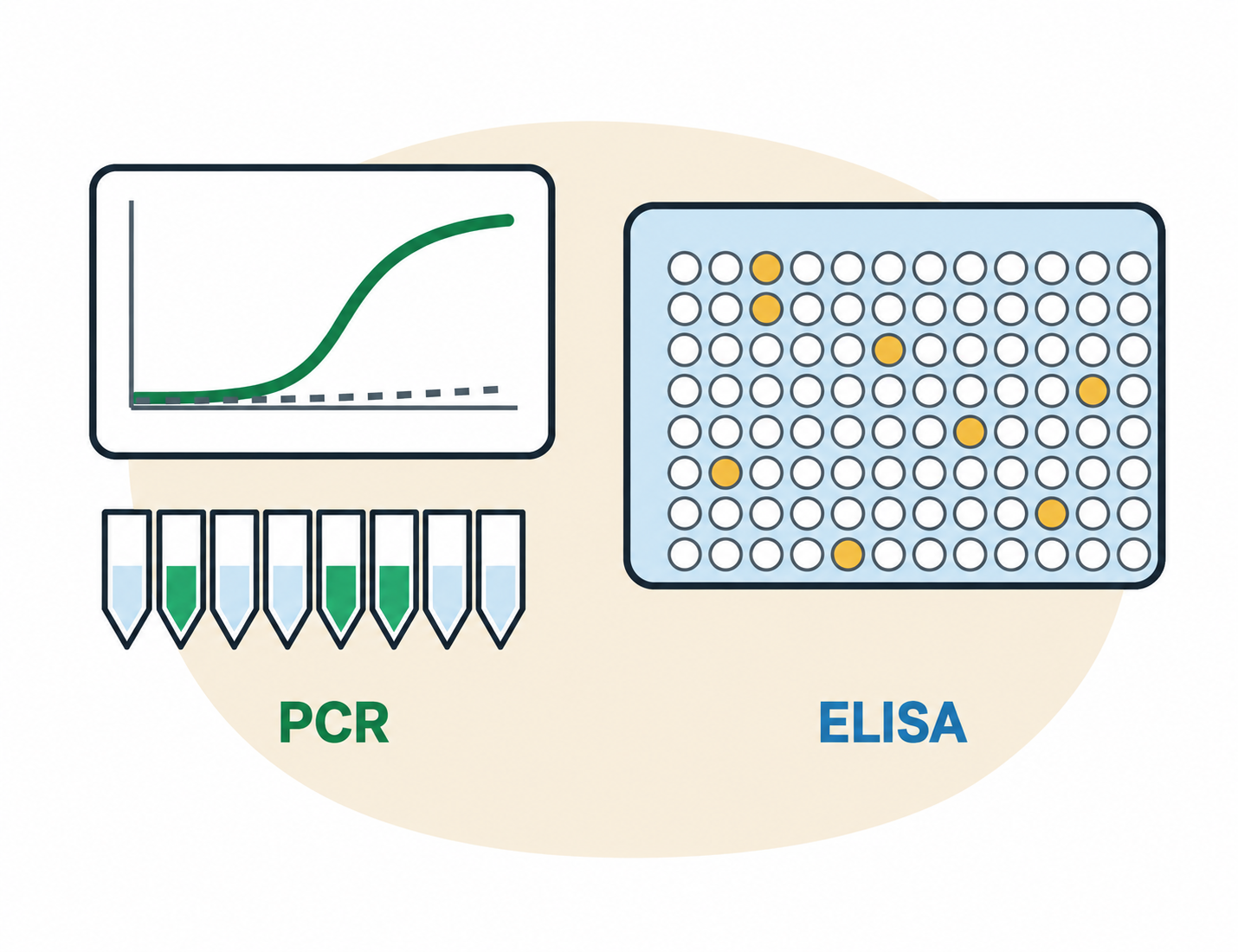
The PCR is looking for the DNA of the virus and confirms the case; Inoculations are recorded among the inoculated animals. The ELISA is looking for an antibodies and shows whether the body has met with a virus or vaccine. Regulation No. 588 provides that antibodies alone are not sufficient for diagnosis. If found, a PCR or a virus study (para. 18).
| Real-time PCR | ELISA | |
|---|---|---|
| What it detects | Viral DNA | Antibodies to capripoxviruses |
| From when | From the onset of fever and skin nodules | According to EFSA, 2 or 4 weeks after the infection; After vaccination from 20 days (producer data) |
| Purpose | Confirm a case; test suspected and contact animals, and semen | Assess post-vaccination antibodies; perform screening and retrospective investigations |
| Limitations | Vaccine virus DNA may be detected after administration of a live vaccine | Does not distinguish vaccinated from recovered animals, or lumpy skin disease virus from sheep pox and goat pox viruses |
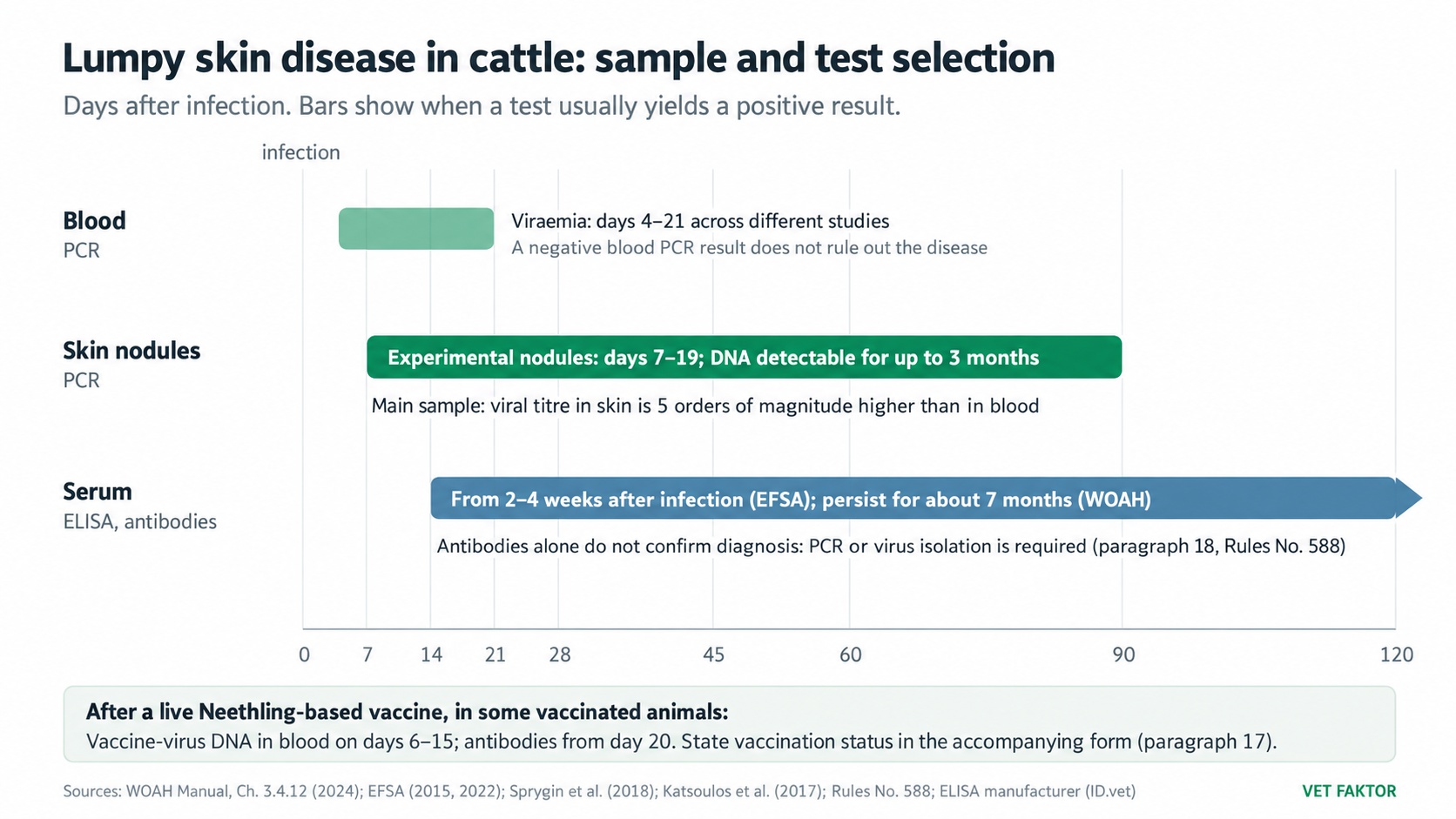
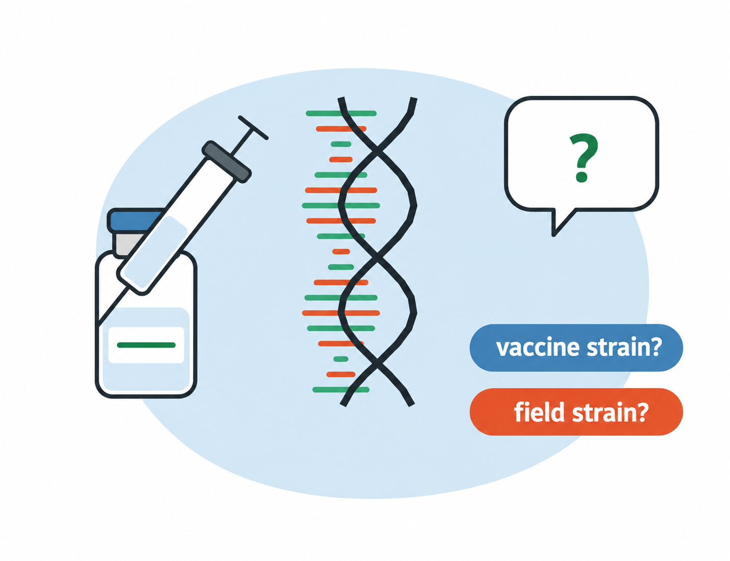
Russia has registered a homologous live vaccine, ZUD vak, based on the Neethling-ARRIAH strain, as well as heterologous vaccines based on sheep pox virus. Following vaccination with a live Neethling strain-based vaccine, the vaccine virus replicates in the animal. In one study, its DNA was detected in blood on days 6–15 in 17 of 27 vaccinated animals. Some animals develop a local reaction at the injection site and, occasionally, skin nodules.
Differentiating PCR assays (DIVA PCR) help distinguish field virus from vaccine virus. They differentiate Neethling vaccine strains from classical field strains. The VET FAKTOR catalogue includes the PCR-LUMPY-SKIN-DISEASE-DIFF-FAKTOR kit: according to the manufacturer, it detects viral DNA and differentiates Neethling strain-based vaccines.
Since 2017, recombinant strains containing genomic regions from both vaccine and field viruses have also been detected in Russia. In areas where they circulate, the WOAH manual recommends whole-genome sequencing to distinguish vaccine from field virus. A differentiating PCR result in a vaccinated animal must therefore be assessed alongside the time since vaccination, clinical findings and the epidemiological situation; sequencing is performed when uncertainty remains. In Russia, such testing is carried out, among others, by the Federal Centre for Animal Health (ARRIAH).
VET FAKTOR specialists can help select kits for your laboratory’s needs and equipment: +7 (495) 789-48-11, vet@faktor.ru.
No. According to the WOAH manual, lumpy skin disease virus is not transmissible to humans. However, the veterinary rules require milk from affected or suspected cows to be destroyed, as well as semen from affected bulls (paragraph 31 of Rules No. 588).
There is no specific antiviral treatment; care is symptomatic. Within an outbreak, affected animals are isolated and treated, while clinically healthy animals are vaccinated (subparagraph b of paragraph 30 of Rules No. 588). Decisions are made by the state veterinary service.
Quarantine is lifted 30 calendar days after the last affected animal in the outbreak has recovered or been slaughtered, and after all other required measures have been completed (paragraph 35). For a further year, unvaccinated susceptible animals may not be introduced into the outbreak site or affected locality. No susceptible animals, including vaccinated ones, may leave the affected locality except for slaughter at slaughterhouses or appropriately equipped slaughter points. Agricultural fairs, exhibitions, auctions and other events bringing susceptible animals together are also prohibited (paragraph 37).
According to the manufacturer of ID Screen® Capripox Double Antigen, antibodies are detectable from day 20 after vaccination. Serum for post-vaccination antibody testing should not be collected earlier. According to the WOAH manual, a negative antibody result in a vaccinated animal does not mean it is unprotected: immunity to capripoxviruses is predominantly cell-mediated, and antibody levels after vaccination may be low.
ELISA detects antibodies, which develop after both vaccination and infection. PCR is needed to confirm a case; in vaccinated animals, its results must be interpreted in the light of vaccination history.
This material is for reference only and does not replace veterinary rules or a veterinary professional’s assessment. Diagnosis is established on the basis of laboratory findings in accordance with the procedure prescribed by law.


© 2015–2026 VET FAKTOR LLC. All rights reserved.